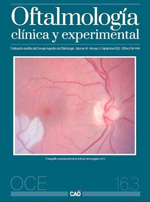
Impacto da hipertensão arterial sistêmica e do diabetes mellitus na espessura da coroide e na microvasculatura da retina por meio da tomografia de coerência óptica
DOI:
https://doi.org/10.70313/2718.7446.v16.n03.241Palavras-chave:
coroide, diabetes mellitus, hipertensão arterial, tomografia de coerência óptica, OCT-AResumo
Objetivos: Determinar se existe relação entre a espessura da coroide e a vascularização superficial da retina com a presença de diabetes mellitus ou hipertensão arterial sistêmica utilizando a técnica EDI-OCT e OCT-A como ferramenta de avaliação.
Materiais e métodos: Estudo transversal observacional, prospectivo e analítico de pacientes saudáveis com diabetes e/ou hipertensão arterial submetidos, como parte da consulta oftalmológica, a EDI-OCT para medição da espessura da coróide e OCT-A para medição da vascularização e a perfusão retiniana.
Resultados: Foram avaliados 168 pacientes com idade de 52,48 ± 13,6 anos, dos quais 57% (96) eram saudáveis, 40,5% (67) apresentavam diabetes mellitus e 47,7% (32) apresentavam hipertensão arterial sistêmica associada. Foi demonstrado que em idade mais avançada ocorre uma diminuição da espessura da coróide de 257,7 ± 59,43 μm (p<0,001). A espessura da coroide subfoveal foi 246,58 ± 55,74 μm no diabetes (p=0,02), espessura da coroide nasal 233,22 ± 54,77 μm (p=0,02), temporal 237,24 ± 57,55 μm (p=0,02). Ao analisar o OCT-A, a densidade vascular periférica em pacientes saudáveis foi de 20,99 ± 2,25 mm2, em diabéticos 19,60 ± 2,17 mm2 (p<0,001) e com retinopatia diabética 18,95 ± 2,24 mm2 (p<0,001). A densidade total de vasos em indivíduos saudáveis foi de 19,74 ± 2,24 mm2 (p<0,001) e para diabetes mellitus 18,37 ± 2,16 mm2 (p<0,001). Não houve diferenças significativas entre os grupos na zona avascular foveal.
Conclusão: A espessura da coroide diminui não só com a idade dos pacientes, mas também no diabetes mellitus, na hipertensão arterial sistêmica e naqueles com ambas as patologias. A densidade dos vasos retinianos e a perfusão vascular são menores em pacientes diabéticos com retinopatia diabética e edema macular diabético do que em pacientes do grupo controle.
Downloads
Referências
Magliano DJ, Boyko EJ. IDF diabetes atlas [en línea]. Brussels: International Diabetes Federation (IDF), 2021. Disponible en: https://www.ncbi.nlm.nih.gov/books/NBK581934/
Singh SR, Vupparaboina KK, Goud A et al. Choroidal imaging biomarkers. Surv Ophthalmol 2019; 64: 312-333.
Pichi F, Aggarwal K, Neri P et al. Choroidal biomarkers. Indian J Ophthalmol 2018; 66: 1716-1726.
Sorrentino FS, Matteini S, Bonifazzi C et al. Diabetic retinopathy and endothelin system: microangiopathy versus endothelial dysfunction. Eye (Lond) 2018; 32: 1157-1163.
Sivaprasad S, Gupta B, Crosby-Nwaobi R, Evans J. Prevalence of diabetic retinopathy in various ethnic groups: a worldwide perspective. Surv Ophthalmol 2012; 57: 347-370.
Abadia B, Suñen I, Calvo P et al. Choroidal thickness measured using swept-source optical coherence tomography is reduced in patients with type 2 diabetes. PLoS One 2018; 13: e0191977.
Rayess N, Rahimy E, Ying GS et al. Baseline choroidal thickness as a predictor for response to anti-vascular endothelial growth factor therapy in diabetic macular edema. Am J Ophthalmol 2015; 159: 85-91.
Lee MW, Koo HM, Lee WH et al. Impacts of systemic hypertension on the macular microvasculature in diabetic patients without clinical diabetic retinopathy. Invest Ophthalmol Vis Sci 2021; 62: 21.
Lim HB, Lee MW, Park JH et al. Changes in ganglion cell-inner plexiform layer thickness and retinal microvasculature in hypertension: an optical coherence tomography angiography study. Am J Ophthalmol 2019; 199: 167-176.
Laviers H, Zambarakji H. Enhanced depth imaging-OCT of the choroid: a review of the current literature. Graefes Arch Clin Exp Ophthalmol 2014; 252: 1871-1883.
Ramrattan RS, van der Schaft TL, Mooy CM et al. Morphometric analysis of Bruch’s membrane, the choriocapillaris, and the choroid in aging. Invest Ophthalmol Vis Sci 35: 2857-2864.
Querques G, Lattanzio R, Querques L et al. Enhanced depth imaging optical coherence tomography in type 2 diabetes. Invest Ophthalmol Vis Sci 2012; 53: 6017-6024.
Nagaoka T, Kitaya N, Sugawara R et al. Alteration of choroidal circulation in the foveal region in patients with type 2 diabetes. Br J Ophthalmol 2004; 88: 1060-1063.
McLeod DS, Lutty GA. High-resolution histologic analysis of the human choroidal vasculature. Invest Ophthalmol Vis Sci 1994; 35: 3799-3811.
Abalem MF, Veloso HNS, Garcia R et al. The effect of glycemia on choroidal thickness in different stages of diabetic retinopathy. Ophthalmic Res 2020; 63: 474-482.
Kinoshita T, Imaizumi H, Shimizu M et al. Systemic and ocular determinants of choroidal structures on optical coherence tomography of eyes with diabetes and diabetic retinopathy. Sci Rep 2019; 9: 16228.
American Academy of Ophthalmology. Retina and vitreous 2020/2021. San Francisco, USA: AAO, 2021: p. 91-93. (Basic and clinical science course; 12).
Mangeaud A, Panigo E. R-Medic: un programa de análisis estadísticos sencillo e intuitivo. Methodo 2018; 3: 18-22. Disponible en: https://methodo.ucc.edu.ar/files/vol3/num1/05%20Methodo%202018_03_01%20Bioestadística%20y%20Metodologia%20aplicada%202018_03_01%20R-medic%20Mangeaud%20A%20et%20al.pdf
Shao L, Zhou LX, Xu L, Wei WB. The relationship between subfoveal choroidal thickness and hypertensive retinopathy. Sci Rep 2021; 11: 5460.
Sun Z, Tang F, Wong R et al. OCT angiography metrics predict progression of diabetic retinopathy and development of diabetic macular edema: a prospective study. Ophthalmology 2019; 126: 1675-1684.
Shin YI, Nam KY, Lee WH et al. Peripapillary microvascular changes in patients with systemic hypertension: an optical coherence tomography angiography study. Sci Rep 2020; 10: 6541.
Waghamare S, Mittal S, Pathania M et al. Comparison of choroidal thickness in systemic hypertensive subjects with healthy individuals by spectral domain optical coherence tomography. Indian J Ophthalmol 2020; 69: 1183-1188
Downloads
Publicado
Edição
Secção
Licença
Direitos de Autor (c) 2023 Consejo Argentino de Oftalmología

Este trabalho encontra-se publicado com a Licença Internacional Creative Commons Atribuição-NãoComercial-SemDerivações 4.0.
Con esta licencia no se permite un uso comercial de la obra original, ni la generación de obras derivadas. Las licencias Creative Commons permiten a los autores compartir y liberar sus obras en forma legal y segura.


