
Bear’s path
congenital hypertrophy of the retinal pigment epithelium
DOI:
https://doi.org/10.70313/2718.7446.v18.n2.420Keywords:
congenital hypertrophy, retinal pigment epithelium, retinal pigmented lesions, choroidal melanoma, congenital hamartoma, familial adenomatous poliposis, colo-rectal tumorAbstract
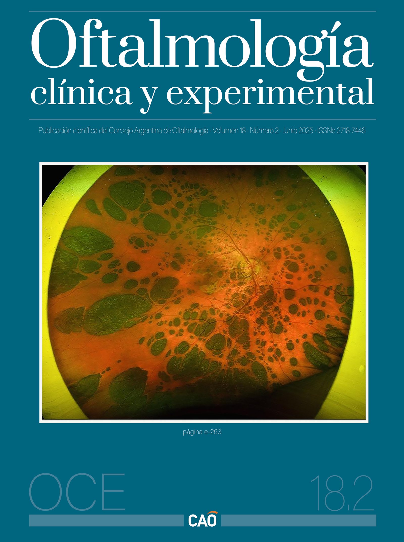
Congenital hypertrophy of the retinal pigment epithelium (CHRPE) is of unknown etiology, manifesting as a pigmented fundus lesion (dark brown to black), flat and well demarcated, typically benign, asymptomatic, stable and generally non-progressive, located at the level of the retinal pigment epithelium (RPE)1. In younger patients (10 to 20 years) the RPE appears homogeneously black and in older individuals there are gaps of depigmentation2. When several lesions of different sizes are joined together they resemble the footprint of an animal and are also called “bear print”3. The main differential diagnosis is with choroidal malignant melanoma, the main difference is in the demarcation of the CHRPE with respect to melanoma and that the malignant lesions are elevated, with not well demarcated borders and can grow in 3 dimensions1-2.
It is a congenital hamartoma of the retinal pigment epithelium (RPE) and occurs in three variants: solitary (unifocal), clustered (multifocal) and atypical1. Atypical CHRPE is associated with familial adenomatous polyposis (FAP), an autosomal dominant cancerous syndrome characterized by numerous adenomatous polyps in the colon and rectum4. If left untreated, virtually all patients with FAP develop colorectal carcinoma in middle age5. Subtypes of FAP, such as Gardner syndrome (FAP plus skeletal hamartomas and various soft tissue tumors) and Turcot syndrome (FAP plus various brain tumors), are also associated with atypical CHRPE 5. In relatives of patients with Gardner’s syndrome, visualization of CHRPE serves as confirmation of the disease before other extraocular tumors appear1-2, 5.
The two figures show multiple bilateral rounded, flat, darkly pigmented, rounded, multiple lesions with flat borders, covering the entire fundus, forming a “bear print” pattern. The patient was a 9 year old girl in whom alterations were detected in the fundus compatible with an CHRPE. The finding was explained to the parents and a follow-up control was recommended in the next six months.
Downloads
References
1. Gallego-Pinazo R, Cotino JF, Dolz-Marco R. Hipertrofia congénita del epitelio pigmentario de la retina. Arch Soc Esp Oftalmol 2016; 91(8): e78. doi:10.1016/j.oftal.2016.02.018.
2. Braga CS, Ricardo OMP, Cordeiro FM, Vieira JM, Nogueira FB. Suspect asymptomatic lesions: congenital hypertrophy of the retinal pigment epithelium (CHRPE). Rom J Ophthalmol 2021; 65(3): 275-278. doi:10.22336/rjo.2021.55.
3. Marmoy OR, Blackwell C, Cornelius S, Thompson DA, Henderson RH. Diffuse bear-track retina: profound, bilateral, grouped congenital pigmentation of the retinal pigment epithelium in an infant. J AAPOS 2020; 24(6): 384-386. doi:10.1016/j.jaapos.2020.08.003.
4. Bonnet LA, Conway RM, Lim LA. Congenital hypertrophy of the retinal pigment epithelium (CHRPE) as a screening marker for familial adenomatous polyposis (FAP): systematic literature review and screening recommendations. Clin Ophthalmol 2022; 16: 765-774. doi:10.2147/OPTH.S354761.
5. Deibert B, Ferris L, Sanchez N, Weishaar P. The link between colon cancer and congenital hypertrophy of the retinal pigment epithelium (CHRPE). Am J Ophthalmol Case Rep 2019; 15: 100524. doi:10.1016/j.ajoc.2019.100524.
Downloads
Published
Issue
Section
License
Copyright (c) 2025 Consejo Argentino de Oftalmología

This work is licensed under a Creative Commons Attribution-NonCommercial-NoDerivatives 4.0 International License.
Con esta licencia no se permite un uso comercial de la obra original, ni la generación de obras derivadas. Las licencias Creative Commons permiten a los autores compartir y liberar sus obras en forma legal y segura.
How to Cite


