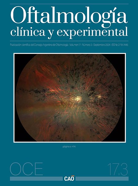
Resultados visuales, refractivos y estructurales a largo plazo en bebés prematuros tratados por retinopatía del prematuro
DOI:
https://doi.org/10.70313/2718.7446.v17.n03.343Palabras clave:
retinopatía del prematuro, fotocoagulación con láser, inyección intravítrea antiangiogénica, miopíaResumen
Objetivo: Analizar la agudeza visual mejor corregida, el estado refractivo y los resultados anatómicos a largo plazo en bebés prematuros tratados consecutivamente por ROP mediante fotocoagulación con láser y/o inyecciones intravítreas de agentes antiangiogénicos.
Métodos: Estudio analítico retrospectivo. Se incluyeron aquellos niños tratados por ROP desde 1998 mediante fotocoagulación con láser, inyecciones intravítreas de agentes antiangiogénicos, o ambos, y que tuvieron un seguimiento oftalmológico mínimo de tres años postratamiento. Se utilizó estadística descriptiva y de frecuencia, y para asociación estadística se utilizó la prueba de Chi-cuadrado y la prueba de correlación de Spearman.
Resultados: Se estudiaron 123 ojos de 62 pacientes. La edad en el último control fue en promedio 9,66 ± 5,61 años. 116 ojos (94%) fueron tratados mediante fotocoagulación con láser, 5 (4%) ojos requirieron tratamiento combinado y 2 (2%) ojos con inyección intravítrea de antiangiogénicos. El equivalente esférico medio fue de -2,68±4,87 dioptrías, siendo significativamente más negativo en aquellos con tratamiento combinado (-11,54±2,36 D) y retratamiento (-12,46±3,26 D) y en aquellos con enfermedad de la zona posterior (-7,50±5,49 frente a +0,52±0,54 D) (p<0,01). Dieciséis (15%) ojos eran emétropes, 32 (29%) ojos eran hipermétropes y 62 (57%) ojos eran miopes. 82 (74%) ojos tenían astigmatismo y 20 (37%) pacientes tenían anisometropía. Sesenta (60%) ojos tenían buena agudeza visual, 26 (25%) ojos tenían agudeza visual regular y 16 (15%) tenían agudeza visual mala. En 97 (80%) ojos el fondo de ojo no presentó secuelas de la enfermedad ni de otras patologías.
Conclusiones: La mayoría de los niños tratados conservan una buena agudeza visual, a pesar de importantes errores refractivos, principalmente miopía. Cuanto más posterior es la enfermedad, mayor es la prevalencia de miopía alta.
Citas
Gupta A, Tripathy K. Central serous chorioretinopathy. En: StatPearls [en línea]. Treasure Island (FL): StatPearls Publishing, 2023 Aug. Disponible en: https://www.ncbi.nlm.nih.gov/books/NBK558973/
Fung AT, Yang Y, Kam AW. Central serous chorioretinopathy: a review. Clin Exp Ophthalmol 2023; 51: 243-270.
Park JB, Kim K, Kang MS et al. Central serous chorioretinopathy: treatment. Taiwan J Ophthalmol 2022; 12: 394-408.
Ivanišević M, Stanić R, Ivanišević P, Vuković A. Albrecht von Graefe (1828-1870) and his contributions to the development of ophthalmology. Int Ophthalmol 2020; 40: 1029-1033.
Rosen E. Central serous retinopathy. Am J Ophthalmol 1948; 31: 734.
Bennett G. Central serous retinopathy. Br J Ophthalmol 1955; 39: 605-618.
Kitzmann AS, Pulido JS, Diehl NN et al. The incidence of central serous chorioretinopathy in Olmsted County, Minnesota, 1980-2002. Ophthalmology 2008; 115: 169-173.
Facello Olmedo FM, Ormaechea G. Coriorretinopatía serosa central aguda y crónica: cambios coroideos observados con tomografía de coherencia óptica con imagen de profundidad mejorada. Oftalmol Clin Exp 2021; 14: 71-80.
Daruich A, Matet A, Dirani A et al. Central serous chorioretinopathy: recent findings and new physiopathology hypothesis. Prog Retin Eye Res 2015; 48: 82-118.
Daruich A, Matet A, Marchionno L et al. Acute central serous chorioretinopathy: factors influencing episode duration. Retina 2017; 37: 1905-1915.
Ficker L, Vafidis G, While A, Leaver P. Long-term follow-up of a prospective trial of argon laser photocoagulation in the treatment of central serous retinopathy. Br JOphthalmol 1988; 72: 829-834.
Gerendas BS, Kroisamer JS, Buehl W et al. Correlation between morphological characteristics in spectral-domain-optical coherence tomography, different functional tests and a patient's subjective handicap in acute central serous chorioretinopathy. Acta Ophthalmol 2018; 96: e776-e782.
Wang M, Munch IC, Hasler PW et al. Central serous chorioretinopathy. Acta
Ophthalmol 2008; 86: 126-145.
Yannuzzi NA, Mrejen S, Capuano V et al. A central hyporeflective subretinal lucency correlates with a region of focal leakage on fluorescein angiography in eyes with central serous chorioretinopathy. Ophthalmic Surg Lasers Imaging Retina 2015; 46: 832-836.
Iida T, Yannuzzi LA, Spaide RF et al. Cystoid macular degeneration in chronic central serous chorioretinopathy. Retina 2003; 23: 1-7, quiz 137-138.
Cardillo Piccolino F, Lupidi M, Cagini C et al. Choroidal vascular reactivity in central serous chorioretinopathy. Invest Ophthalmol Vis Sci 2018; 59: 3897-3905.
Sahoo NK, Mishra SB, Iovino C et al. Optical coherence tomography angiography findings in cystoid macular degeneration associated with central serous chorioretinopathy. Br J Ophthalmol 2019; 103: 1615-1618.
Mrejen S, Balaratnasingam C, Kaden TR et al. Long-term visual outcomes and causes of vision loss in chronic central serous chorioretinopathy. Ophthalmology 2019; 126: 576-588.
Spaide RF, Klancnik JM Jr. Fundus autofluorescence and central serous chorioretinopathy. Ophthalmology 2005; 112: 825-833.
Conrad R, Geiser F, Kleiman A et al. Temperament and character personality profile and illness-related stress in central serous chorioretinopathy. ScientificWorldJournal 2014; 2014: 631687.
Miki A, Kondo N, Yanagisawa S et al. Common variants in the complement factor H gene confer genetic susceptibility to central serous chorioretinopathy. Ophthalmology 2014; 121: 1067-1072.
Negi A, Marmor M.F. Experimental serous retinal detachment and focal pigment epithelial damage. Arch Ophthalmol 1984; 102: 445-449.
Nicholson B, Noble J, Forooghian F, Meyerle C. Central serous chorioretinopathy: update on pathophysiology and treatment. Surv Ophthalmol 2013; 58: 103-126.
Spaide RF. Choroidal blood flow: review and potential explanation for the choroidal venous anatomy including the vortex vein system. Retina 2020; 40: 1851-1864.
Pang CE, Shah VP, Sarraf D, Freund KB. Ultra-widefield imaging with autofluorescence and indocyanine green angiography in central serous chorioretinopathy. Am J Ophthalmol 2014; 158: 362.e2-371.e2.
Brinks J, van Dijk EHC, Meijer OC et al. Choroidal arteriovenous anastomoses: a hypothesis for the pathogenesis of central serous chorioretinopathy and other pachychoroid disease spectrum abnormalities. Acta Ophthalmol 2022; 100: 946-959.
Imanaga N, Terao N, Nakamine S et al. Scleral thickness in central serous chorioretinopathy. Ophthalmol Retina 2021; 5: 285-291.
Fernández-Vigo JI, Moreno-Morillo FJ, Shi H et al. Assessment of the anterior scleral thickness in central serous chorioretinopathy patients by optical coherence tomography. Jpn J Ophthalmol 2021; 65: 769-776.
Sirakaya E, Duru Z, Kuçuk B, Duru N. Monocyte to high-density lipoprotein and neutrophil-to-lymphocyte ratios in patients with acute central serous chorioretinopathy. Indian J Ophthalmol 2020; 68: 854-858.
Zola M, Gobeaux C, Javorsky T et al. Galectin 3 and central serous chorioretinopathy: a promising new biomarker. Proc ARVO Annual Meeting 2021; 2021: 2197.
Bahadorani S, Maclean K, Wannamaker K et al. Treatment of central serous chorioretinopathy with topical NSAIDs. Clin Ophthalmol 2019; 13: 1543-1548.
Lehmann M, Bousquet E, Beydoun T, Behar-Cohen F. Pachychoroid: an inherited condition? Retina 2015; 35: 10-16.
Schubert C, Pryds A, Zeng S et al. Cadherin 5 is regulated by corticosteroids and associated with central serous chorioretinopathy. Hum Mutat 2014; 35: 859-867.
Zhang X, Lim CZF, Chhablani J, Wong YM. Central serous chorioretinopathy: updates in the pathogenesis, diagnosis and therapeutic strategies. Eye Vis (Lond) 2023; 10: 33.
Potsaid B, Baumann B, Huang D et al. Ultrahigh speed 1050 nm swept source/fourier domain OCT retinal and anterior segment imaging at 100,000 to 400,000 axial scans per second. Opt Express 2010; 18: 20029-20048.
Warrow DJ, Hoang QV, Freund KB. Pachychoroid pigment epitheliopathy. Retina 2013; 33: 1659-1672.
Gal-Or O, Dansingani KK, Sebrow D et al. Inner choroidal flow signal attenuation in pachychoroid disease: optical coherence tomography angiography. Retina 2018; 38: 1984-1992.
Burumcek E, Mudun A, Karacorlu S, Arslan MO. Laser photocoagulation for persistent central serous retinopathy: results of long-term follow-up. Ophthalmology 1997; 104: 616-622.
Roca JA, Wu L, Fromow-Guerra J et al. Yellow (577 nm) micropulse laser versus half-dose verteporfin photodynamic therapy in eyes with chronic central serous chorioretinopathy: results of the Pan-American Collaborative Retina Study (PACORES) Group. Br J Ophthalmol 2018; 102: 1696-1700.
Zeng M, Chen X, Song Y, Cai C. Subthreshold micropulse laser photocoagulation versus half-dose photodynamic therapy for acute central serous chorioretinopathy. BMC Ophthalmol 2022; 22: 110.
Yannuzzi LA, Slakter JS, Gross NE et al. Indocyanine green angiography-guided photodynamic therapy for treatment of chronic central serous chorioretinopathy: a pilot study. Retina 2003; 23: 288-298.
van Dijk EHC, Fauser S, Breukink MB et al. Half-dose photodynamic therapy versus high-density subthreshold micropulse laser treatment in patients with chronic central serous chorioretinopathy: the place trial. Ophthalmology 2018; 125: 1547-1555.
Manayath GJ, Narendran V, Arora S et al. Graded subthreshold transpupillary thermotherapy for chronic central serous chorioretinopathy. Ophthalmic Surg Lasers Imaging 2012; 43: 284-290.
Peiretti E, Caminiti G, Serra R et al. Anti-vascular endothelial growth factor therapy versus photodynamic therapy in the treatment of choroidal neovascularization secondary to central serous chorioretinopathy. Retina 2018; 38: 1526-1532.
Duan J, Zhang Y, Zhang M. Efficacy and safety of the mineralocorticoid receptor antagonist treatment for central serous chorioretinopathy: a systematic review and meta-analysis. Eye (Lond) 2021; 35: 1102-1110.
Daugirdas SP, Bheemidi AR, Singh RP. Should we stop treating patients with eplerenone for chronic CSCR?: commentary on the VICI trial. Ophthalmic Surg Lasers Imaging Retina 2021; 52: 308-310.
Feenstra HMA, van Dijk EHC, van Rijssen TJ et al. Long-term follow-up of chronic central serous chorioretinopathy patients after primary treatment of oral eplerenone or half-dose photodynamic therapy and crossover treatment: SPECTRA trial report No. 3. Graefes Arch Clin Exp Ophthalmol 2023; 261: 659-668.
Salehi M, Wenick AS, Law HA et al. Interventions for central serous chorioretinopathy: a network meta-analysis. Cochrane Database Syst Rev 2015; 2015: CD011841.
Gramajo AL, Marquez GE, Torres VE et al. Therapeutic benefit of melatonin in refractory central serous
horioretinopathy. Eye (Lond) 2015; 29: 1036-1045.
Lai TYY, Staurenghi G, Minerva Study Group et al. Efficacy and safety of ranibizumab for the treatment of choroidal neovascularization due to uncommon cause: twelve-month results of the Minerva Study. Retina 2018; 38: 1464-1477.
Descargas
Publicado
Cómo citar
Número
Sección
Licencia
Derechos de autor 2024 Consejo Argentino de Oftalmología

Esta obra está bajo una licencia internacional Creative Commons Atribución-NoComercial-SinDerivadas 4.0.
Con esta licencia no se permite un uso comercial de la obra original, ni la generación de obras derivadas. Las licencias Creative Commons permiten a los autores compartir y liberar sus obras en forma legal y segura.


