
Ceratite bacteriana difusa em pacientes com ceratopatia bolhosa
DOI:
https://doi.org/10.70313/2718.7446.v17.n02.331Palavras-chave:
ceratopatia bolhosa, ceratite infecciosa, ceratite bacteriana, colírios fortificadosResumo
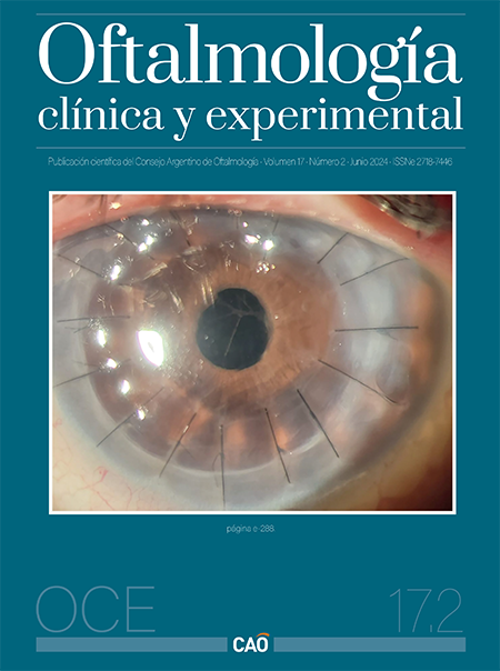
A descompensação endotelial da córnea pode ter diferentes causas. Sua manifestação clínica provoca um quadro denominado ceratite bolhosa em que se observa edema corneano com perda de transparência e alteração estrutural que se manifesta por ciclos recorrentes de lesões bolhosas no epitélio corneano. O objetivo deste trabalho é relatar uma série de casos de pacientes com ceratopatia bolhosa que apresentaram ceratite infecciosa corneana cujo principal sinal era o hipópio. Após coleta de amostra para exame direto e cultura, foi realizado tratamento com colírios fortificados (vancomicina 50 mg/ml e ceftazidima 50 mg/ml), obtendo resposta satisfatória no contexto de infecções bacterianas. Enfatiza-se considerar a ceratopatia bolhosa como fator de risco para ceratite infecciosa, onde pode haver infecção bacteriana da córnea sem ulceração ou infiltração evidente no momento, e onde a presença de hipópio é o único sinal de infecção da córnea nos casos apresentados.
Downloads
Referências
Austin A, Lietman T, Rose-Nussbaumer J. Update on the management of infectious keratitis. Ophthalmology 2017; 124: 1678-1689.
Ljubimov AV, Atilano SR, Garner MH et al. Extracellular matrix and Na+, K+-ATPase in human corneas following cataract surgery: comparison with bullous keratopathy and Fuchs’ dystrophy corneas. Cornea 2002; 21: 74-80.
Gomes JAP, Haraguchi DKM, Zambrano DU et al. Punções do estroma anterior no tratamento da ceratopatia bolhosa. Arq Bras Oftalmol 2000; 63: 133-137.
Wollensak G, Green WR, Seiler T. Corneal myxoma. Jpn J Ophthalmol 2002; 46: 193-197.
Morishige N, Sonoda KH. Bullous keratopathy as a progressive disease: evidence from clinical and laboratory imaging studies. Cornea 2013; 32 Suppl 1: S77-S83.
Gonçalves ED, Campos M, Paris F et al. Ceratopatia bolhosa: etiopatogênese e tratamento. Arq Bras Oftalmol 2008; 71 (6 Suppl): 61-64.
Eaton JS, Miller PE, Bentley E et al. The SPOTS system: an ocular scoring system optimized for use in modern preclinical drug development and toxicology. J Ocul Pharmacol Ther 2017; 33: 718-734.
Ung L, Bispo PJM, Shanbhag SS et al. The persistent dilemma of microbial keratitis: global burden, diagnosis, and antimicrobial resistance. Surv Ophthalmol 2019; 64: 255-271.
Kaye SB, Rao PG, Smith G et al. Simplifying collection of corneal specimens in cases of suspected bacterial keratitis. J Clin Microbiol 2003; 41: 3192-3197.
Grandi G, Bianco G, Boattini et al. Bacterial etiology and antimicrobial resistance trends in ocular infections: a 30- year study, Turin area, Italy. Eur J Ophtalmol 2021; 31: 405-414.
Acharya M, Farooqui JH, Gaba T et al. Delhi infectious keratitis study: update on clínico-microbiological profile and outcomes of infectious keratitis. J Curr Ophtalmol 2020; 32: 249-255.
Kansky JJ. Oftalmología clínica. Barcelona: Elseiver España, 2016.
Fernández Ferreiro A, González-Barcia M, Gil-Martínez M et al. Evaluation of the in vitro ocular toxicity of the fortified antibiotic eye drops prepared at the Hospital Pharmacy Departments. Farm Hosp 2016; 40: 352-370.
Siu GD, Young AL, Jhanji V. Alternatives to corneal transplantation for the management of bullous keratopathy. Curr Opin Ophthalmol 2014; 25: 347-352.
Singh M, Sinha BP, Mishra D et al. Role of corneal collagen cross-linking in bullous keratopathy: a systematic review. Indian J Ophthalmol 2023; 71: 1706-1717.
Downloads
Publicado
Edição
Secção
Licença
Direitos de Autor (c) 2024 Consejo Argentino de Oftalmología

Este trabalho encontra-se publicado com a Licença Internacional Creative Commons Atribuição-NãoComercial-SemDerivações 4.0.
Con esta licencia no se permite un uso comercial de la obra original, ni la generación de obras derivadas. Las licencias Creative Commons permiten a los autores compartir y liberar sus obras en forma legal y segura.
Como Citar


