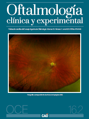
Pit de papila associada a maculopatia e descolamento seroso da retina
apresentação de dois casos
DOI:
https://doi.org/10.70313/2718.7446.v16.n02.234Palavras-chave:
pit de papila, fosseta, fosseta de papila, descolamento seroso da retina, vitrectomiaResumo
Objetivo: Apresentar os casos de dois pacientes com hipófise e descolamento seroso da retina associado, patologia raramente relatada na literatura.
Caso clínico: Caso 1: Homem de 47 anos que consultou por diminuição súbita da acuidade visual unilateral. Foi diagnosticado como coriorretinopatia serosa central. Após vários ciclos de tratamento habitual sem boa resposta, chegou-se ao diagnóstico de papila pituitária com descolamento seroso da retina associado. Caso 2: Paciente do sexo feminino, 38 anos, que consultou devido a uma diminuição súbita da acuidade visual unilateral. Foi inicialmente diagnosticado como uma complicação de um evento isquêmico retiniano. Após ter realizado tratamento sem resposta satisfatória, chegou-se ao diagnóstico de papila hipofisária com descolamento seroso de retina associado. A resolução em ambos os casos foi cirúrgica por meio de vitrectomia com injeção de gás, endofotocoagulação e peeling da membrana limitante interna, com resultados anatômicos favoráveis.
Conclusão: O descolamento de retina associado à fosseta de papila representa um desafio diagnóstico. De acordo com a experiência adquirida e a bibliografia científica publicada, há necessidade de ferramentas que facilitem o diagnóstico precoce para melhorar o prognóstico visual dos pacientes.
Downloads
Referências
Ferry AP. Macular detachment associated with congenital pit of the optic nerve head: pathologic findings in two cases simulating malignant melanoma of the choroid. Arch Ophthalmol 1963;70: 346-357.
Regenbogen L, Stein R, Lazar M. Macular and juxtapapillar serous retinal detachment associated with pit of optic disc. Ophthalmologica 1964; 148: 247-251.
Meng L, Zhao X, Zhang W et al. The characteristics of optic disc pit maculopathy and the efficacy of vitrectomy: a systematic review and meta-analysis. Acta Ophthalmol 2021; 99: e1176-e1189.
Wan R, Chang A. Optic disc pit maculopathy: a review of diagnosis and treatment. Clin Exp Optom 2020; 103: 425-429.
Georgalas I, Ladas I, Georgopoulos G, Petrou P. Optic disc pit: a review. Graefes Arch Clin Exp Ophthalmol 2011; 249: 1113-1122.
Brodsky MC. Congenital optic disk anomalies. Surv Ophthalmol 1994; 39: 89-112.
Van Dijk EHC, Boon CJF. Serous business: delineating the broad spectrum of diseases with subretinal fluid in the macula. Prog Retin Eye Res 2021; 84: 100955.
Moisseiev E, Moisseiev J, Loewenstein A. Optic disc pit maculopathy: when and how to treat?: a review of the pathogenesis and treatment options. Int J Retina Vitreous 2015; 1: 13.
Sugar HS. An explanation for the acquired macular pathology associated with congenital pits of the optic disc. Am J Ophthalmol 1964; 57: 833-835.
Sugar HS. Congenital pits in the optic disc with acquired macular pathology. Am J Ophthalmol 1962; 53: 307-311.
Tavallali A, Sadeghi Y, Abtahi SH et al. Inverted ILM flap technique in optic disc pit maculopathy. J Ophthalmic Vis Res 2023 19; 18: 230-239.
Christoforidis JB, Terrell W, Davidorf FH. Histopathology of optic nerve pit-associated maculopathy. Clin Ophthalmol 2012; 6: 1169-1174.
Regenbogen L, Stein R, Lazar M. Macular and juxtapapillar serous retinal detachment associated with pit of optic disc. Ophthalmologica 1964; 148: 247-251.
Kuhn F, Kover F, Szabo I, Mester V. Intracranial migration of silicone oil from an eye with optic pit. Graefes Arch Clin Exp Ophthalmol 2006; 244: 1360-1362.
Sobol WM, Blodi CF, Folk JC, Weingeist TA. Long-term visual outcome in patients with optic nerve pit and serous retinal detachment of the macula. Ophthalmology 1990; 97: 1539-1542.
Oli A, Balakrishnan D. Treatment outcomes of optic disc pit maculopathy over two decades. Ther Adv Ophthalmol 2021; 13: 25158414211027715.
Pinheiro RL, Henriques F, Figueira J et al. Surgical approaches to optic disc pit maculopathy: a clinical case series. Case Rep Ophthalmol 2022; 13: 885-891.
Slocumb RW, Johnson MW. Premature closure of inner retinal fenestration in the treatment of optic disk pit maculopathy. Retin Cases Brief Rep 2010; 4: 37-39.
Gass JD. Serous detachment of the macula: secondary to congenital pit of the optic nervehead. Am J Ophthalmol 1969; 67: 821-841.
Cox MS, Witherspoon CD, Morris RE, Flynn HW. Evolving techniques in the treatment of macular detachment caused by optic nerve pits. Ophthalmology 1988; 95: 889-896.
Lee KJ, Peyman GA. Surgical management of retinal detachment associated with optic nerve pit. Int Ophthalmol 1993; 17: 105-107.
Chatziralli I, Theodossiadis P, Theodossiadis GP. Optic disk pit maculopathy: current management strategies. Clin Ophthalmol 2018; 12: 1417-1422.
Downloads
Publicado
Edição
Secção
Licença
Direitos de Autor (c) 2023 Consejo Argentino de Oftalmología

Este trabalho encontra-se publicado com a Licença Internacional Creative Commons Atribuição-NãoComercial-SemDerivações 4.0.
Con esta licencia no se permite un uso comercial de la obra original, ni la generación de obras derivadas. Las licencias Creative Commons permiten a los autores compartir y liberar sus obras en forma legal y segura.


