Retinal and spinal cord involvement in pheochromocytoma
a diagnostic challenge
DOI:
https://doi.org/10.70313/2718.7446.v16.n02.233Keywords:
pheochromocytoma, hypertensive retinopathy, macular star, optic disc edemaAbstract
Objective: To present a clinical case of pheochromocytoma with retinal and spinal cord involvement, and to review the relevance of the interdisciplinary approach for its diagnosis and treatment.
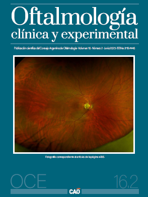
Clinical case: A 15-year-old woman consulted for a sudden decrease in visual acuity and headaches of 3 months of evolution. Bilateral papillae edema was observed and magnetic resonance imaging of the brain with suspected intracranial hypertension was requested. Hyperintense area at the bulbomedullary junction was detected, complemented with cervical resonance imaging, obtaining findings compatible with demyelinating disease, establishing a probable diagnosis of neuromyelitis optica. In his evolution he presented severe arterial hypertension. The case was re-evaluated and observed in background of retinal involvement (bilateral macular star). The picture is reinterpreted as hypertensive retinopathy and secondary causes are sought. In a new interrogation, antecedents compatible with pheochromocytoma were highlighted: “headaches, palpitations and sweating”. Ultrasound and abomen MRI detected lesion and high values of catecholamines were detected in urine and serum confirming the diagnosis. The BP was controlled and two weeks later the tumor was operated and removed.
Conclusion: The diagnosis of pheochromocytoma can be dificult. An interdisciplinary approach is required to assess the different clinical expressions and also to carry out the corresponding treatment.
References
Neumann HPH, Young WF Jr, Eng C. Pheochromocytoma and paraganglioma. N Engl J Med 2019; 381: 552-565.
Farrugia FA, Martikos G, Tzanetis P et al. Pheochromocytoma, diagnosis and treatment: review of the literature. Endocr Regul 2017; 51: 168-181.
Farrugia FA, Charalampopoulos A. Pheochromocytoma. Endocr Regul 2019; 53: 191-212.
Younes S, Abdellaoui M, Zahir F et al. Bilateral stellate neuroretinitis revealing a pheochromocytoma. Pan Afr Med J 2015; 20: 13.
Carrasquillo JA, Chen CC, Jha A et al. Imaging of pheochromocytoma and paraganglioma. J Nucl Med 2021; 62: 1033-1042.
Takahashi K. Adrenomedullin from a pheochromocytoma to the eye: implications of the adrenomedullin research for endocrinology in the 21st century. Tohoku J Exp Med 2001; 193: 79-114.
Guilmette J, Sadow PM. A guide to pheochromocytomas and paragangliomas. Surg Pathol Clin 2019; 12: 951-965.
Lee AG, Beaver HA. Acute bilateral optic disk edema with a macular star figure in a 12-year-old girl. Surv Ophthalmol 2002; 47: 42-49.
Gocmen R, Ardicli D, Erarslan Y et al. Reversible hypertensive myelopathy-the spinal cord variant of posterior reversible encephalopathy syndrome. Neuropediatrics 2017; 48: 115-118.
McKinney AM, Short J, Truwit CL et al. Posterior reversible encephalopathy syndrome: incidence of atypical regions of involvement and imaging findings. AJR Am J Roentgenol 2007; 189: 904-912.
Published
How to Cite
Issue
Section
License
Copyright (c) 2023 Consejo Argentino de Oftalmología

This work is licensed under a Creative Commons Attribution-NonCommercial-NoDerivatives 4.0 International License.
Con esta licencia no se permite un uso comercial de la obra original, ni la generación de obras derivadas. Las licencias Creative Commons permiten a los autores compartir y liberar sus obras en forma legal y segura.


