SCIENTIFIC OPINIONS
“The eye knows”: hyperspectral images
Luciana L. Iacono
Hospital de Clínicas José de San Martín, Buenos Aires, Argentina.
Received: April 24th, 2023.
Approved: May 19th, 2023.
Corresponsal author
Luciana L. Iacono MD
Córdoba 2351
(C1028) Buenos Aires, Argentina.
+54 11 5950-8000
lucianaliacono@gmail.com
Oftalmol Clin Exp (ISSNe 1851-2658)
2023; 16(2): e99-e102.
Acknowledgments
To Dr. Andrea Barral, for establishing the contacts that allowed me to learn about this new technological development.
To the engineer Denis Hellebuyck, CEO of Mantis Photonics (Lund, Sweden), for providing the product information.
Abstract
As ophthalmologists we know that the eye, besides being part of the visual system, is an organ where multiple systemic pathologies can be expressed both in their initial stages and in their evolution. But new technologies, with which a large amount of data can be get and related, aim to make diagnoses from the eye at much earlier stages. Research activities are showing us this future, as in the case of hyperspectral imaging. Currently under development are devices that can get hyperspectral images of the retina by means of which early stages of ocular, cardiovascular and neurodegenerative diseases could be detected.
Keywords: hyperspectral imaging, retina, neurodegeneration, Alzheimer’s disease.
“El ojo sabe”: imágenes hiperespectrales
Resumen
Como médicos oftalmólogos sabemos que el ojo, además de formar parte del sistema visual, es un órgano donde se pueden expresar múltiples patologías sistémicas tanto en sus etapas iniciales como en su evolución. Pero nuevas tecnologías con las que se pueden obtener y relacionar gran cantidad de datos pretenden realizar diagnósticos a partir del ojo en etapas mucho más tempranas. Las actividades de investigación nos muestran ese futuro, como es el caso de las imágenes hiperespectrales. Actualmente están en desarrollo unos dispositivos que pueden conseguir imágenes hiperespectrales de la retina mediante la cual se podrían detectar estadios precoces de patologías oculares, cardiovasculares y enfermedades neurodegenerativas.
Palabras clave: imágenes hiperespectrales, retina, neurodegeneración, enfermedad de Alzheimer.
“O olho sabe”: imagens hiperespectrais
Resumo
Como oftalmologistas sabemos que o olho, além de fazer parte do sistema visual, é um órgão onde múltiplas patologias sistêmicas podem se manifestar tanto em seus estágios iniciais quanto em sua evolução. Mas novas tecnologias com as quais grandes quantidades de dados podem ser obtidas e relacionadas visam fazer diagnósticos do olho em estágios muito anteriores. As atividades de pesquisa nos mostram esse futuro, como é o caso das imagens hiperespectrais. Atualmente, estão sendo desenvolvidos dispositivos que podem obter imagens hiperespectrais da retina, através das quais podem ser detectados estágios iniciais de patologias oculares, cardiovasculares e doenças neurodegenerativas.
Palavras-chave: imagens hiperespectrais, retina, neurodegeneração, doença de Alzheimer.
That “the retina is the window to the central nervous system” we have already heard more than once but that “the eye is the window to aging” could be considered a new and upcoming reality, which shows us hyperspectral images.
What is hyperspectral imaging?
The spectrum of information classically obtained from digital images of the retina is limited, since only monochromatic or trichromatic light (red, green, blue) reflected by retinal structures is recorded1. Hyperspectral imaging technology has the potential to overcome changes in retinal reflectance and light absorbance that occur during the development of various eye diseases, by producing an image that captures information from multiple wavelengths, generating a four-dimensional hyperspectral cube: two dimensions for orthogonal spatial data, a third for wavelength bands and finally, one for their corresponding absorbance/reflectance intensities at each wavelength1-2. Hyperspectral imaging collects data from tens to hundreds of narrow-wavelength bands, while multispectral imaging refers to data 3 to 10 bands wider1.
Medical applications of hyperspectral imaging: general aspects
Although this type of information is being evaluated for application in different areas of medicine1, a main interest has been generated in ophthalmology, since it is a specialty whose diagnoses depend largely on the acquisition and analysis of the images obtained2-3. For example, an innovative hyperspectral retinal camera for the detection of multiple systemic pathologies is currently under development. This does not refer to a new software applied to the optical coherence tomography (OCT) but to a new instant camera, capable of identifying multiple wavelengths (Fig. 1)4. It is a device that is under development by a spin-off company from Lund University, Sweden, called Mantis Photonics AB4. One of its founders, engineer Denis Hellebuyck, refers to the new device as portable, easy to use, low cost and generator of a high quality hyperspectral image (information obtained from your website)5. And as we have proven in scientific publications, the analysis of hyperspectral images could allow us to identify ocular pathologies (glaucoma, age-related macular degeneration), cardiovascular diseases and neurodegenerative diseases such as Alzheimer’s disease in early stages of the disease2-3, 6. Likewise, this type of technology could be very interesting for the follow-up of treatments or interventions aimed at slowing down the cellular aging process, although for that, there is a long way to go.

Figure 1. Sequence of 6 images of the optic papilla and an adjacent area at different wavelengths, taken with a hyperfractal camera from Mantis Photonics (images courtesy of Mantis Photonics, Lund, Sweden).
Hyperspectral imaging in diabetic retinopathy and age-related macular degeneration
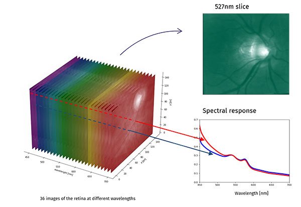
This type of device, developed to take hyperspectral images, could quantify the oxygenation of the retina, so it would be possible to objectively monitor hypoxia (Fig. 2) in patients with cardiovascular pathologies and especially in people with diabetic retinopathy or vascular occlusions, among other alterations2-3. In fact, this type of technology is already in use and evaluation in other areas, for diabetic foot monitoring7. But this technology could also measure and analyze the contour of macular drusen to monitor age-related macular degeneration3 and measure the contour and type of nevi and/or pigmented lesions in the fundus6.

Figure 2. The 36-wavelength image generated with the Mantis Photonics camera. The analysis of the optical density spectrum allow to determine, among other things, the oxygenation of the retinal tissue (image courtesy of Mantis Photonics, Lund, Sweden).
Hyperspectral imaging and Alzheimer’s disease
One of the most potential qualities of this new technology is oriented to the neurological level, for the early detection of Alzheimer’s disease and other neurodegenerative dementias, quantitatively measuring amyloid and/or other neurological biomarkers at the retinal level8-10. It is estimated that amyloid would be present at this level several years prior to the onset of the patient’s cognitive disorder, so it is even speculated that the diagnosis could be established 10 to 15 years prior to the appearance of clinical symptoms10. But although the vertiginous advances of science excite us about the near future, confirmatory scientific data will be necessary, which in turn must be validated over time.
In conclusion, in a not too distant future, new medical devices for ophthalmological use could take advantage of the information obtained from the detailed analysis of the retina, and as we have seen particularly from hyperspectral imaging, which are already a reality. Through these images obtain structural information (quantitative and qualitative) and functional biochemistry, which would be useful for the detection and early treatment not only of eye diseases, but also of neurological and cardiovascular pathologies. Ophthalmology associated with the great scientific-technological growth is increasingly relevant in the context of general health and vascular and neurodegenerative pathologies in particular.
References