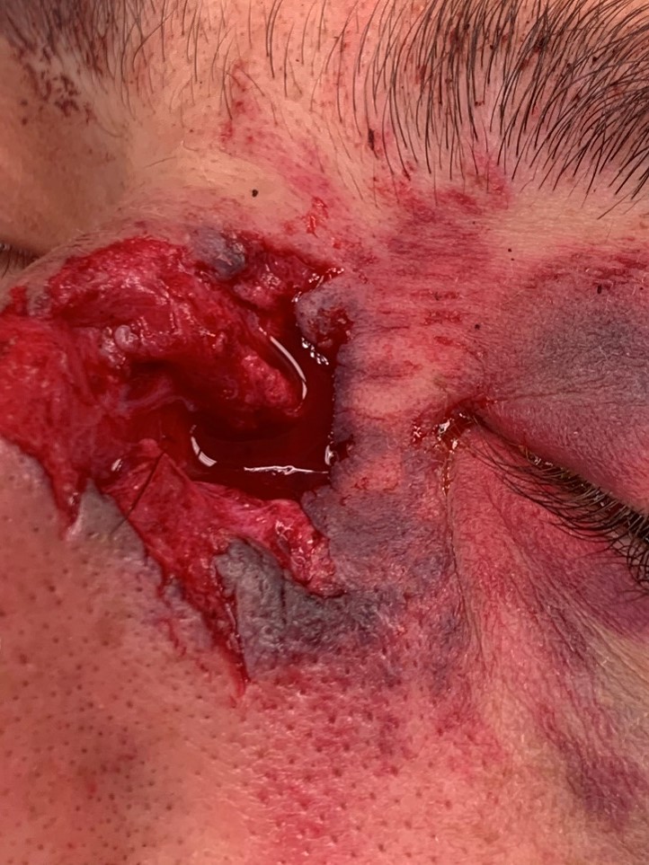
Figura 1. Orificio de entrada.
IMÁGENES CIENTÍFICAS
Una herida de bala y mucha suerte
Jorge Sánchez-Monroy
Departamento de Oftalmología, Hospital Universitario Miguel Servet, Zaragoza, España.
Departamento de Cirugía, Universidad de Zaragoza, España.
Recibido: 18 de julio de 2021.
Aceptado: 14 de octubre de 2021.
Autor corresponsal
Dr. Jorge Sánchez-Monroy
Departamento de Oftalmología
Hospital Miguel Servet
Paseo Isabel la Católica, 1-3
50009 Zaragoza, España.
jrgsanchez.cr@gmail.com
Oftalmol Clin Exp (ISSN 2718-7446)
2022; 15(1): e90-e91.
Conflicto de intereses
El autor declara que no tener conflicto de intereses.
Las heridas de bala orbitarias son potencialmente severas, su extracción es compleja y el pronóstico visual generalmente malo1-3. En las imágenes del presente caso se puede apreciar el orificio de entrada de la bala (fig. 1) y su localización final a nivel orbitario (fig. 2). El proyectil se alojó inferior al nervio óptico en su salida del globo; el trayecto orbitario fue limpio sin lesionar estructuras. Se realizó la extracción del proyectil en órbita izquierda mediante abordaje orbitario a través de incisión subciliar. Se observó hematoma en recto inferior y se constató la integridad del globo. No se encontró ningún signo de lesión oftalmológica tras la cirugía y se preservó la visión en todo momento, a pesar de la relevancia del evento sufrido y las severas consecuencias que podrían haber acontecido.
A bullet wound with a happy end
Orbital gunshot wounds are potentially severe, their extraction is complex and the visual prognosis is generally poor1-3. In the images of the present case, the entry hole of the bullet can be seen (fig. 1) and its final location at the orbital level (fig. 2). The projectile was lodged inferior to the optic nerve at its exit from the globe, the orbital path was clean without damaging structures. The projectile was extracted in the left orbit by means of an orbital approach through a subciliary incision. Hematoma was produced in the lower rectum and the integrity of the globe was verified. No signs of ophthalmological injury were found after surgery and vision was preserved at all times, despite the relevance of the event suffered and the serious consequences that could have occurred.
Figura 1. Orificio de entrada.

Figura 2. Visualización de la bala en la cavidad orbitaria mediante imagen TAC.
Referencias
1. Promsopa C, Prapaisit U. Removal of intraconal bullet through endoscopic transnasal surgery with image-guided navigation system 8 months after injury: a case report. J Med Case Rep 2019; 13: 65.
2. Cho SM, Cho D, Read-Fuller A, Vrcek I. Orbitozygomatic approach for bullet retrieval from the temporal fossa. Proc (Bayl Univ Med Cent) 2019; 32: 435-437.
3. Elegbede A, Drogt C, Wasicek PJ et al. Orbital injuries from self-inflicted gunshots: patterns, management, and visual outcomes. Ophthalmic Plast Reconstr Surg 2020; 36: 152-156.